Bioimaging Core Facility

cellshorizontal.gif
Clipboard.png Clipboard-2.png


The Bioimaging core facility is specialized in light microscopy. Its goal is to help users from experimental design & acquisitions to image processing (treatment and quantification). In addition to the maintenance of the resources of the platform and the users' training, we are also involved in the organization of workshops about ImageJ and other imaging softwares and in several academic courses. The facility can also help you with data presentation for oral presentation or publication (manipulation of file formats, animations, ...).

PREREQUISITE: If you want to use the Bioimaging core facility for the first time (microscopes and/or computers), please SUBMIT THE REGISTRATION FORM AVAILABLE ON LINE (HERE).


Microscopes & PC workstations housed in our facility are available 24/24 hours, 7/7 days for trained users, with consultation, training and support services available from 8.00 a.m. to 5.00 p.m. (Monday through Friday). Please contact directly François or Olivier.

Microscopes and computers' use is not free of charge. Fees are available HERE. Please do not hesitate to contact us if you have any question.

In term of applications, our microscopes allow to study the :

(i) Localization between different proteins or between diverse biological structures (co-localization, structure/ morphology, large image, 3D rendering, correlative microscopy, super-resolution, ...);

(ii) Dynamics of  live cells and the movement of intracellular structures (cell tracking, multi-dimensional time-lapse acquisitions, FRAP experiments,  Ca2+ imaging,  photoactivation, ...);

(iii) Interactions between cells or different proteins (FRET, ...).


Please contact directly Nicolas or Sergei. They will be pleased to help you in your projects. Consultations and support services are available:

  • Only Friday (from 9:00 a.m. to 6:00 p.m.) (for Sergei).
  • From Monday to Friday (from 8:00 a.m. to 5:00 p.m.) (for Nicolas).

In term of applications, our softwares and skills in image treatment allow to quantify automatically a large diversity of images  (co-localization, counting, fluorescence intensity measurements, ...).

Important: before acquiring a large amount of images, it is always recommended to talk with us the best way to make the acquisitions in order to optimize data analysis.

Fees are available HERE.

Your request will be processed as follow :

  •  A first consultation (free of charges) will be organized with the dedicated Bioimaging personnel to determine which method would be the most appropriate to your scientific needs. This will also help us to estimate the time required for its development.
  •  If the estimated time is less than 2 hours, we will issue directly the invoice.
  • If more than 2 hours are needed, a project request must be formally submitted by email to Bioimaging(at)unige.ch. This request must be approved by the Bioimaging board. The submitted project must comply with the platform's rules and will be processed according to the platform priorities. If the project is accepted, the applicant will be notified by email. Then, a quote will be established. Once your PI has accepted the estimate, the work will start. In any case, if the project requires less time than we plan initially, you will be charged only on the scripting hours done. 
  • The time required for one project must not involve the full-time staff of the platform in an abusive manner. In practice, the Bioimaging is a core facility shared by numerous research groups of the Faculty of Medicine and must dealing simultaneously with multiple projects. This work will be done during the official working hours.
  • Priority will be given to internal groups of the Faculty of Medicine.
  • Your project will be processed in order of arrival (ticket system) and according to the number of projects that the Bioimaging service must be brought to deal with. If possible, try to contact us in advance in order to process your request in the best conditions.
  • The time devoted to data analysis can be reduced if the workload due to with the multiple activities of the platform requires it.


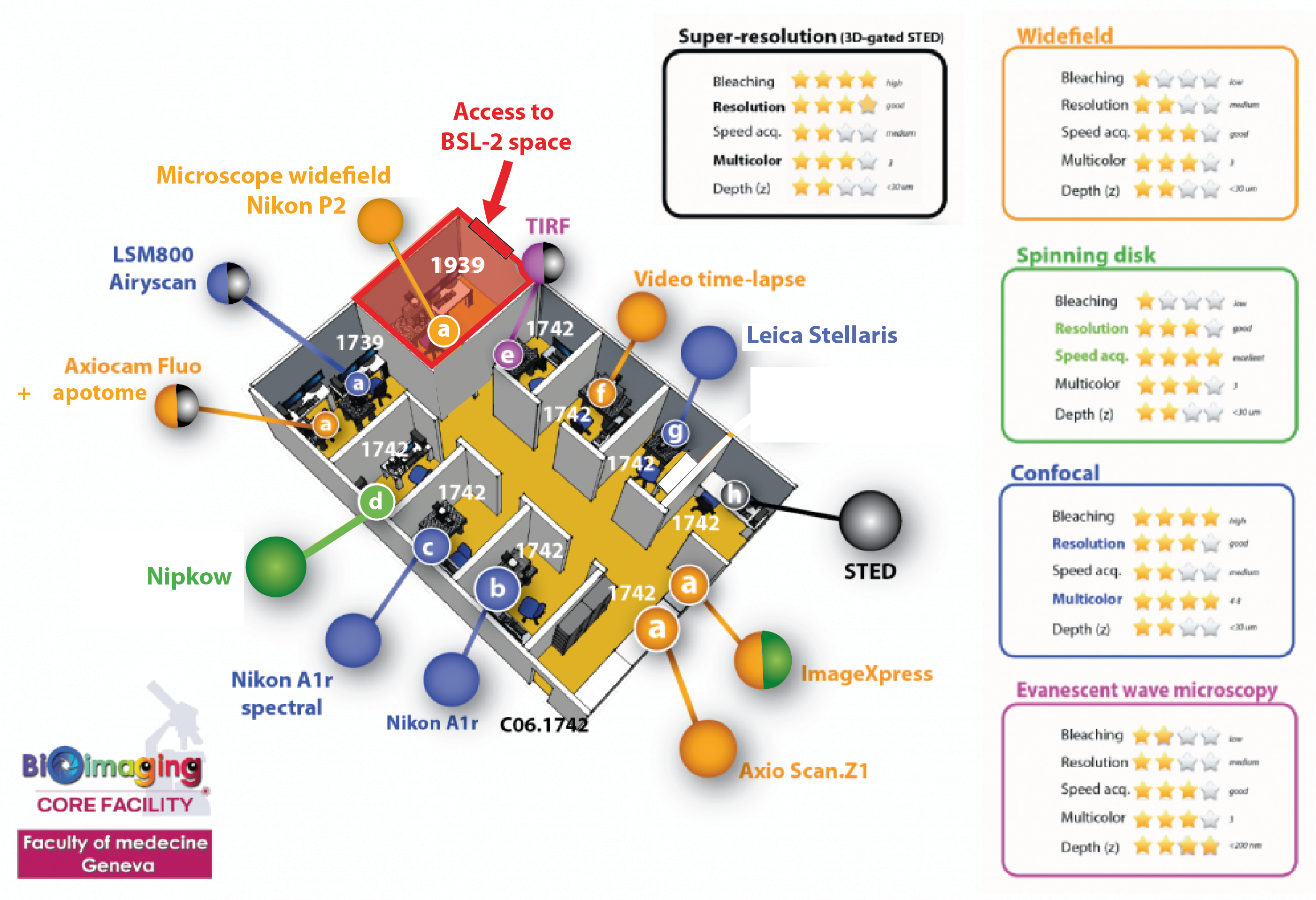
Our bioimaging facility hosts state-of-the-art equipment with:

  • 17 imaging systems (1 STED nanoscope, 1 spinning disk confocal microscope (Nipkow), 5 confocal laser scanning microscopes (CLSM), 2 slide scanners and 8 widefield fluorescence microscopes including 1 TIRF evanescent wave microscope, 1 laser microdissector and 1 plate scanner/ImageXpress system) that provide a wide range of applications such as cell membrane visualization, laser microdissection/isolation, colocalization analysis, F-techniques (FRAP, FRET), multi-dimensional time-lapse recordings, intravital imaging and high-throughput imaging in ROOM C06.1742.a.
  • 7 PC workstations for image processing and analysis (Imaris, Amira, Huygens, metaXpress, MetaMorph, ZEN Lite, NIS, LAS X FLIM, Fiji, Matlab, QuPath, etc...). These PC are located just in front the microscope in ROOM C06.1542.a.

Distribution microscopes.png

The LMD is installed in ROOM D06.1554.a.

The Olympus VS120 is installed in ROOM C06.1533.a.