Ultra-high resolution scanning 3D-EM has arrived at the PFMU
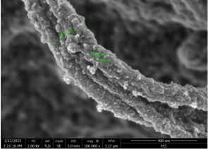
The Electron Microscopy Core Facility (Pôle Facultaire de Microscopie Electronique, PFMU) has recently acquired the latest technology in ultra-structural analysis, the Helios Nanolab G3 scanning 3D-EM microscope from FEI. This over-a-million franc machine that combines the latest in electron imaging, ion beam and robotic technology has an astounding theoretical maximum resolution of 0.7 nm, making it one of the post powerful scanning EM microscopes out there.
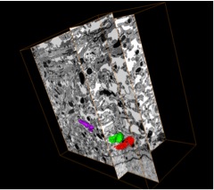
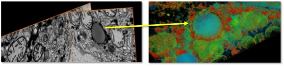
The combination of an ion beam, electron beam and multi-angle motorized stage allows embedded specimens to be imaged and sectioned sequentially, at intervals as low as 3 nm between sections. The collected images can then be later reconstructed into stunning 3D volumes. The catch: one sample might take 2 days of continuous manipulation at the microscope, including an overnight run. Thankfully the software can email you at home to let you know when the imaging is complete!
The funding for this mean machine was raised by a team of UNIGE scientists through an R’Equip grant lead by Prof. Michelangelo Foti and including PHYM members Profs. Pierre Cosson and Nicolas Demaurex.
The Helios microscope in the PFMU core facility will be accessible to all users as of March 2016.
For more information please contact: Michelangelo Foti
Posted by: P. Nunes
13 Dec 2015