Centriole
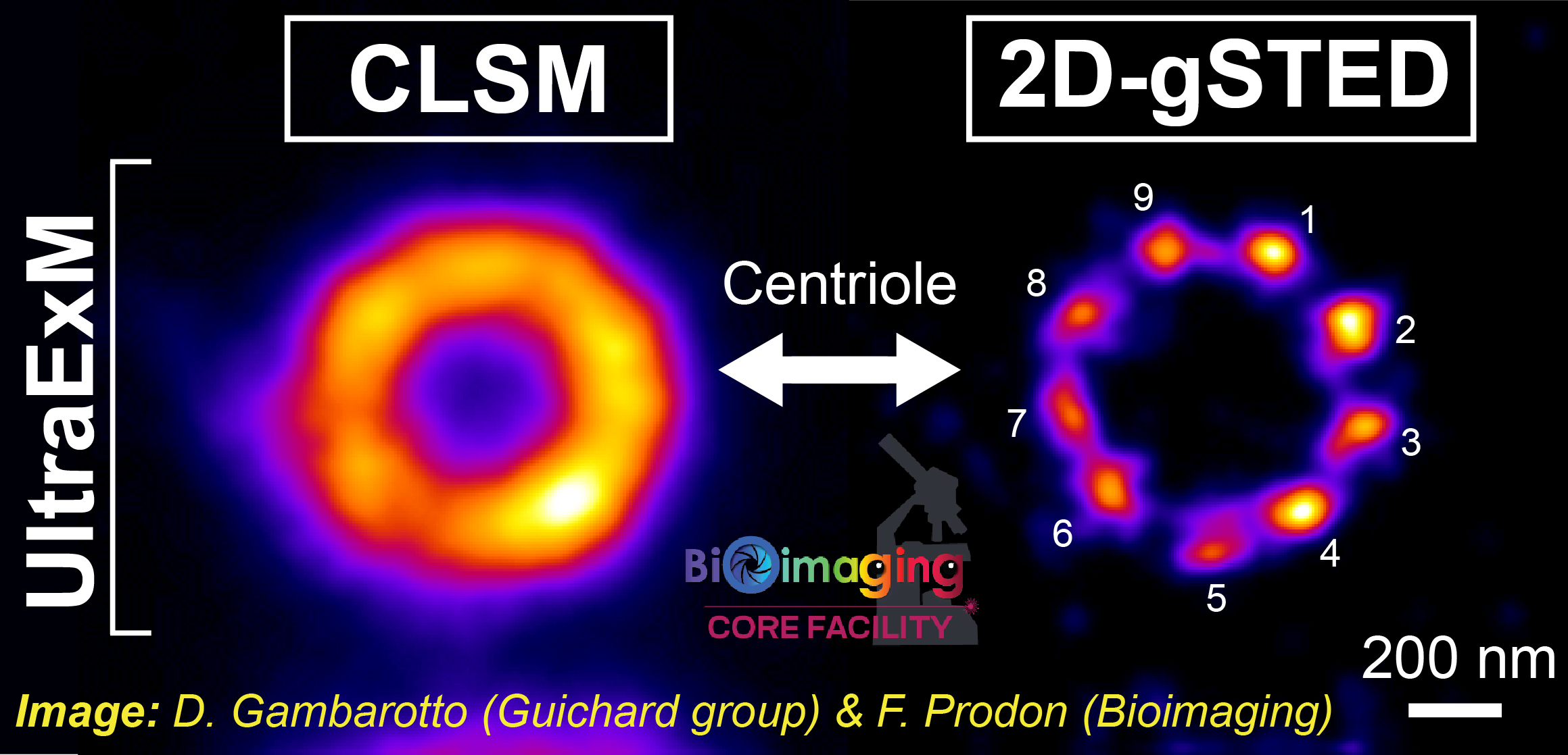
Comparison between confocal microscopy (left) and 2D gated STED nanoscopy (right). The same centriole was observed after having used ultrastructure expansion microscopy (UltraExM). Note the significant gain of spatial resolution...(Image source: © Davide Gambarotto, Guichard lab, Dpt of Cell Biology, Faculty of Sciences & François Prodon, Bioimaging, Faculty of Medicine/CMU ).
Davide's article is available HERE.