Gallery
Deconvolved image of mitotic spindles (microtubules, yellow) and chromosomes (cyan) from a 2-cell stage embryo (C.elegans)
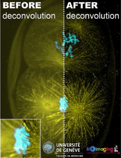
This image corresponds to a Z-projection (from a Z-stack, 100 slices acquired with LSM700 microscope). Huygens software was used for deconvolution with a measured PSF. (Image source:© O. Brun & F. Prodon, Bioimaging Core Facility, Gotta group, CMU)