Protein clusters to better anchor cells
In order to ensure their survival and perform properly their tasks, cells must anchor to their extracellular environment. To do this, they form highly complex protein structures, called focal adhesions. In a recent article in Communications Biology, researchers from the laboratory of Prof. Bernhard Wehrle-Haller have demonstrated the key role of the paxillin protein in the formation of these focal adhesions. However, the spatial organization of paxillin and other proteins within adhesions still remained unclear.
Phosphorylated paxillin is distributed in a periodic pattern like beads on a necklace
The team of researchers from the laboratory of Prof. Bernhard Wehrle-Haller, in collaboration with the team of Prof. Martin Bastmeyer from the Karlsruhe Institute of Technology, were now able to shed light on the localization of the phosphorylated form of paxillin within focal adhesions, a discovery published in the Journal of Cell Science.
Using new super-resolution microscopy techniques, they revealed that phosphorylated paxillinwas not homogeneously distributed in adhesions, but was instead clustered at regular, periodic intervals like beads on a necklace (see images below). In contrast, the entire population of paxillin proteins within adhesions did not show this pattern. The scientists then studied another protein known to interact with only the phosphorylated form of paxillin, FAK, and observed a similar spotty distribution.
Their further analysis of the dynamics of these patterns in living cells suggested that a spontaneous association of the protein FAK with paxillin, might trigger the selective re-distribution of the phosphorylated form of paxillin together with FAK to create more stable adhesive structures.
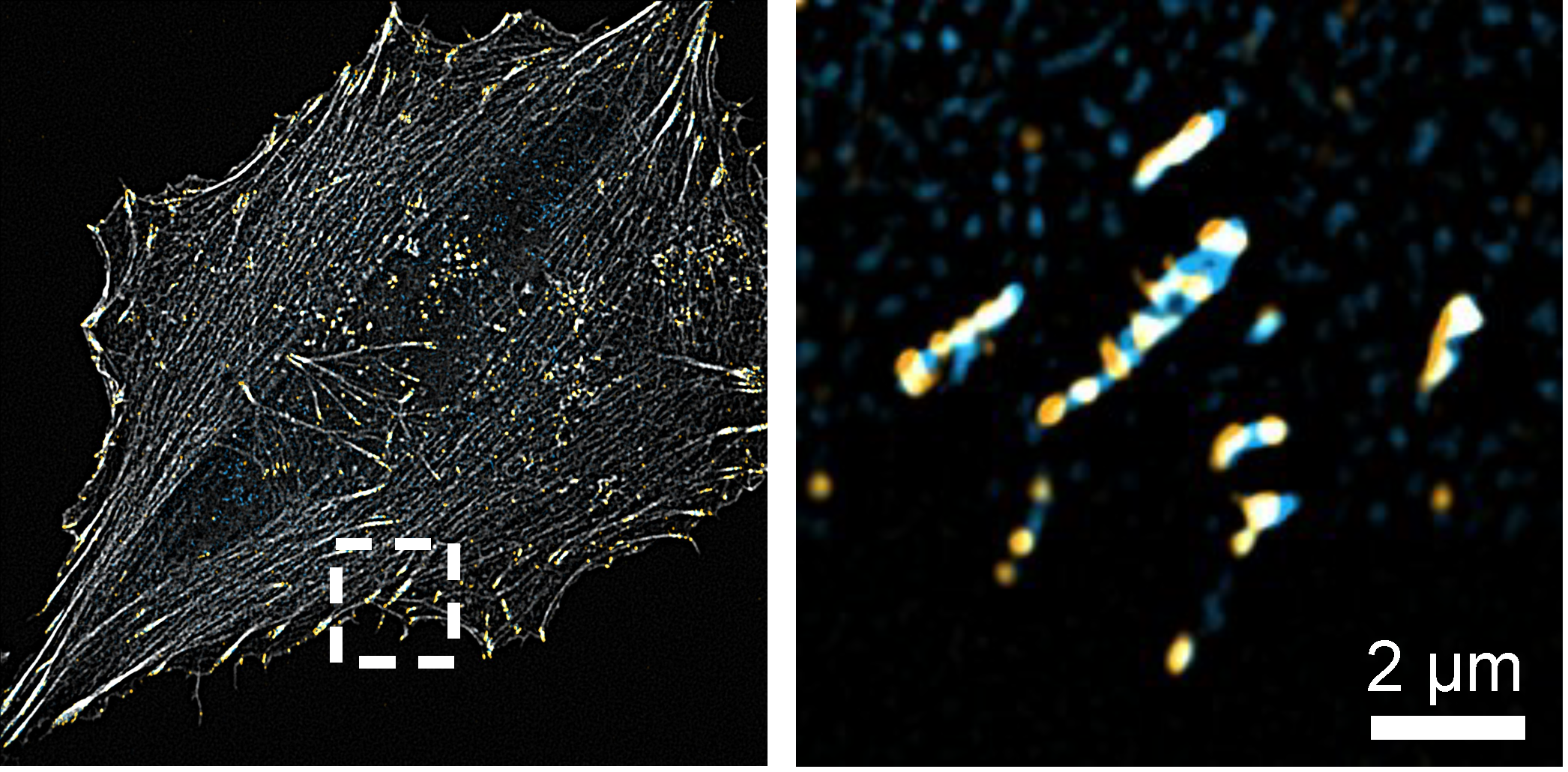
A cell has multiple focal adhesion (left image, in yellow) that connect the cell to its environment. General paxillin (right image, in blue) is distributed throughout focal adhesions, whereas the phosphorylated form of paxillin (right image, in orange) is restricted to distinct points or beads within focal adhesions © Wehrle-Haller laboratory, UNIGE
Potential implications
The regular patterns of phosphorylated paxillin evidenced by the teams of researchers are key elements for cell anchoring and survival. Their findings could therefore help to understand how pathological alterations of cell adhesion processes contribute to cancer progression. The team of Prof. Bernhard Wehrle-Haller plans now to investigate whether other proteins located at focal adhesions show similar patterning and if this patterning is critical for their function.
29 Apr 2022