High Resolution Computed Tomography Imaging (Quantum GX)
In vivo X-Ray Computed Tomography (CT) is a technology that uses X-rays emitted from a focused radiation source and sensed by a detector. The source and detector are rotated around a study subject placed in the middle of the CT scanner. The X-Rays are attenuated at different rates as they pass through different types of tissue. At each emission angle a 2D image is generated that can then be combined into 3D structures by the system's reconstruction software.
The Quantum GX microCT imaging system provides high-resolution images at an X-ray dose low enough to enable true longitudinal imaging capability. The Quantum GX has three modes: high resolution, high speed and standard modes. In the high resolution mode, a 4.5 μm voxel size resolution can be attained at a 36x36 mm FOV, while a 9 μm voxel size resolution can be attained at 72x72 mm FOV.
Quantum GX makes microCT imaging as easy as IVIS and allows you to co-register functional optical signals with anatomical microCT throughout the span of your study. Throughput and user workflow have been simplified to seamlessly co-register anatomical and functional data at every step of a longitudinal study.
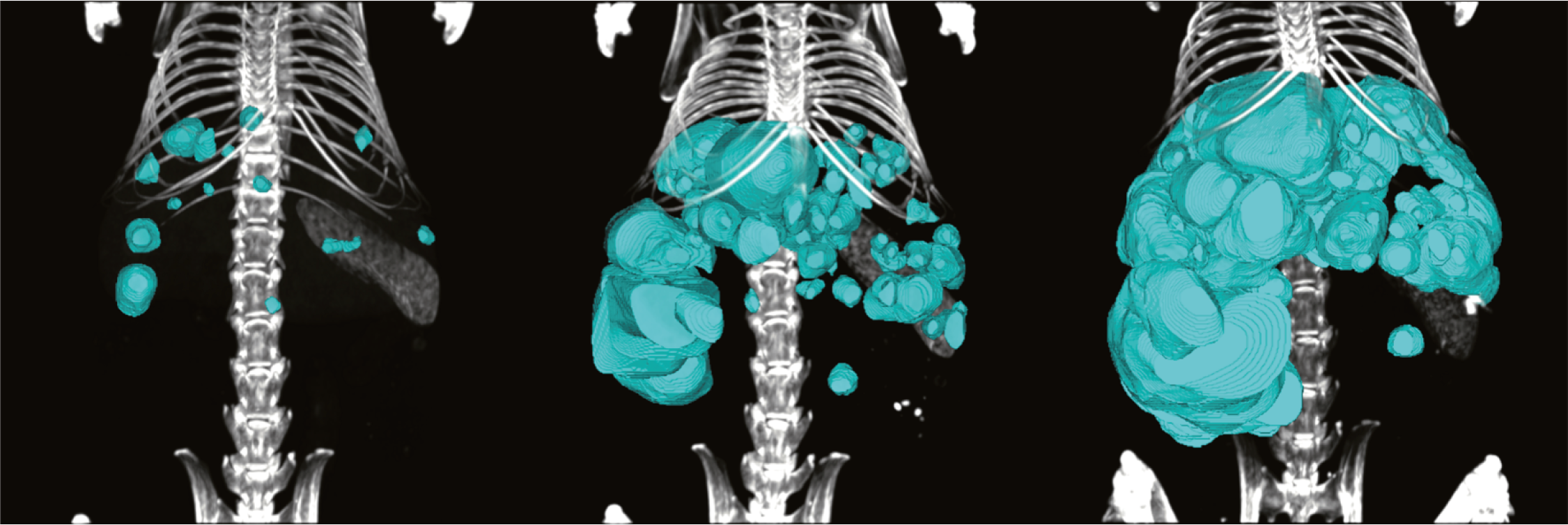
Tumour growth in a liver tumour model at 7, 9 & 11 months
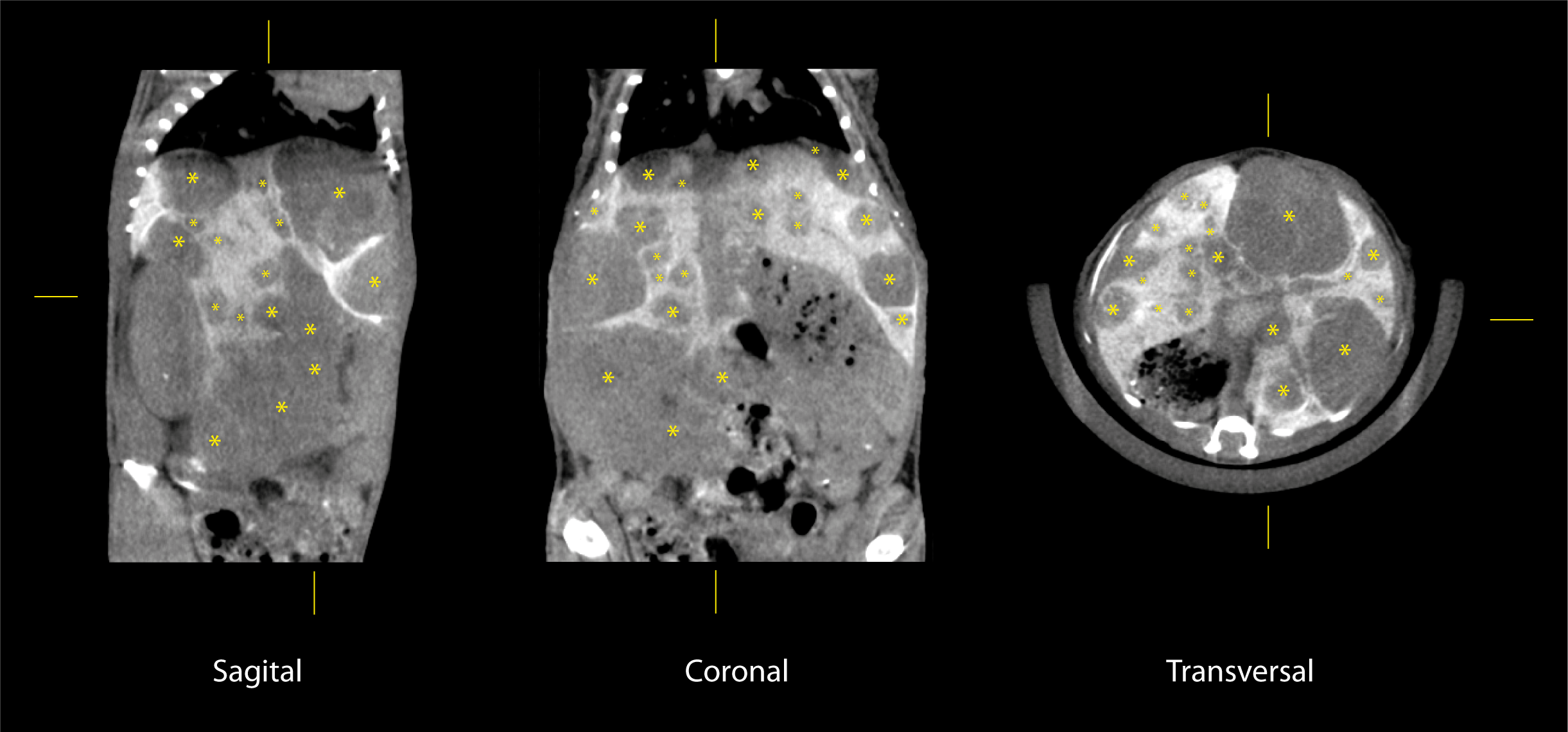
Tumours (yellow asterisk) in a liver tumour model at 11 months

Vascularisation of A) the heart and major vessels, B) the liver, C) the kidneys and D) a tumour

Bone and Joint Destruction in MIA Osteoarthritis Model at 0, 2, 4 & 6 weeks

Co-registration CT (Quantum GX)/Bioluminescence (IVIS)

Segmentation of tumor (blue) bearing lungs (green)
Cardiac gating following vascular contrast agent injection (respiratory gating)
White Adipose tissue segmentation