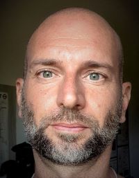
Luigi Bonacina - Coordinator
Biophotonics, University of Geneva
Luigi Bonacina started his academic career at the University of Milan, Italy. After obtaining a Master in Optics, he moved to Switzerland where he completed a PhD at the Swiss Federal Institute of Technology of Lausanne (EPFL) with a research in the field of ultrafast molecular spectroscopy. Successively, he joined the University of Geneva, where he is now Associate Professor at the Department of Applied Physics. His research activities are focused on the development of novel bio-imaging approaches based on the combination of nanotechnology and nonlinear optics. Since 2007, he has been actively working on the development of Harmonic Nanoparticles (HNPs), a family of non-fluorescent dielectric particles emitting multiple harmonic signals by tunable laser excitation from the visible to the Short-Wave Infrared with applications for cell tracking. During his activity, Luigi Bonacina has established a dynamic and truly interdisciplinary network of European researchers as testified by several collaborative publications in the fields of cancer, regenerative, and circadian medicine which go along with articles in the fields of optics, spectroscopy, and nanotechnology.
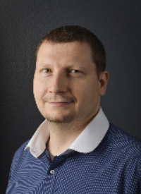
Peter Horvath
Image-based Data Analysis, Szeged Biological Research Centre
Peter Horvath is the Director of the Institute of Biochemistry, Biological Research Centre, BRC, Szeged, Hungary, and the High-Content Analysis Facility, HELMI, Helsinki, Finland. Furthermore, he is a group leader at BRC and was awarded a Finland Distinguished Professor Fellowship at the University of Helsinki (FIMM-EMBL). He obtained his Master's diploma as a Computer Scientist at the University of Szeged, Hungary. In 2008, he finished his PhD at the University of Nice, Sophia Antipolis, France. Since then, he has pioneered the analysis and single cell-based phenotyping of high-content screens for which he developed outstanding image correction, segmentation, and phenotyping methods. Besides, he has participated in data analysis of several of the first dozen whole human genome-wide siRNA screens. He has developed image-based analysis technologies to discover novel drug treatments for cancer patients. Also, he has elaborated the algorithms which have led to the discovery of the role of the HDAC6 gene in and potential drug targets for influenza infection and has developed methods that precisely localize the role of drugs and genes involved in influenza infection. He is the co-founder of the European Cell-based Assays Interest Group (www.eucai.org) and served as a counsellor for the Society of Biomolecular Imaging and Informatics (www.sbi2.org). In general, his groups focus on finding computational solutions to biological problems, combining wet-lab and light microscopy with image analysis and machine learning.
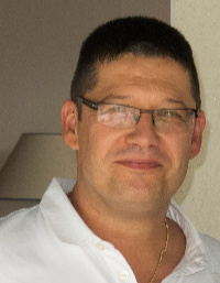
Karl Rouger
Regenerative medicine, INRAE/Oniris PAnTher
Karl Rouger is a senior scientist in cell biology at INRAE/Oniris PAnTher (Animal Pathophysiology & Biotherapy of muscle and nervous system diseases) joint research unit, France. Since 2010, he is deputy director of the unit that integrates a state-of-the-art platform in comparative pathology and fluorescence bio-imaging (APEX), which proposes an integrated offer in tissue and cell phenotyping. After obtaining a Master in physiology and cell biology in 1997, he pursued a PhD in molecular and cell biology at the University of Nantes (France) with research in the field of pathophysiology of muscle diseases (structured around a comparative pathology approach including muscular dystrophies, inflammatory myopathies and vacuolar myopathy). Since end of 2002, he has a permanent position at INRAE in the Animal Health division where he developed a translational research axis devoted to regenerative medicine of muscle diseases with the use of adult stem cells and different animal models. Since 2016, he participates to the Bioregate regenerative medicine cluster and is partner of the Center of reference for rare neuromuscular diseases Atlantique-Occitanie-Caraïbes that participates in different clinical trials in the field of orphan diseases. He has special interest in Myology, diversity of progenitor cells and post-grafting cell fate with exploitation of innovative cell tracking tools. In recent years, he has been particularly involved in understanding the properties and modes of action of adult stem cells with studies on interactions with immune cells as well as on the nature and intensity of muscle tissue remodeling after transplantation.
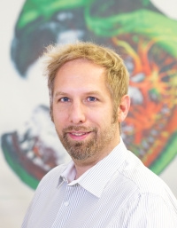
Volker Andresen
Head, LaVision BioTec GmbH
Volker Andresen finished successfully his diploma thesis in 2001 at MPI for Biophysical Chemistry in Göttingen, Dept. of NanoBiophotonics, supervised by Nobel Prize Winner Prof. Dr. S. Hell. He started then his career at LaVision BioTec as Project Manager. In 2003 he became Product manager for Laser-Scanning Microscopy and in 2009 technical director as well as head of department: research, development and production. Since 2013 Volker Andresen is technical director at LaVision BioTec and head of departments: research, strategic cooperations and intellectual property; production; finances. Since 2003 he coordinated different research projects funded by BMBF (MeMo, FLI-Cam, TOMOSphere, ELICIT (with MPG), MoDiaNo) and BMWi (HD-DET, CSIM, LSTM, RETI-IM). Volker Andresen is steakholder of LaVision in Göttingen.
Dirk Theisen-Kunde
Dirk Theisen-Kunde studied Technical Physics at the Polytechnic University Lübeck (Germany) and Physics at the University of Loughborough (UK). After his Diploma-Thesis in “Experimental investigation of plasma formation in water with pico and nanosecond Nd: YAG laser pulses” he worked at the Institute of Biomedical Optics at the University of Lübeck in the field of endoscopic laser surgery. During this time he developed a surgical laser prototype in cooperation with the Bavarian laser company StarMedTec, ending up in a commercial product for surgical laser surgery called Vela Laser.
Since 2010 he is CTO at the Medical Laser Center in Lübeck and is responsible for prototyping of medical and customized laser devices. A “Dental-OCT” Prototype was awarded by the “Dental Innovation Award 2018” in the category “Innovative Idea / Invention”. Together with the R&D team several ophthalmological prototypes were realized for the innovative approach called “Selective Retina Therapy (SRT)” that are used worldwide for scientific evaluation. Also an intra-operative MHz-OCT prototype for neuro-surgery is part of his actual work.
Sebastian "Nino" Karpf
Laser Physicist, University of Lübeck
Sebastian Karpf is a Juniorprofessor for translational biomedical photonics at the Institute of Biomedical Optics (BMO) at the University of Lübeck, Germany. He is the main inventor and driving force behind the SLIDE technology. His expertise spans laser development, fiber optics, electronics, microscopy, imaging, nonlinear optics, programming, data handling and biomedical applications.
Lukas Kontenis
Scientific Laser System, Light Conversion
Lukas Kontenis received his PhD in physics in 2017 from the University of Toronto. During his time at the University of Toronto he worked in the group of Prof. Virginijus Barzda on the topic of biomedical polarimetric harmonic-generation microscopy. He also holds an MSc degree in physics from Vilnius University, where he worked on femtosecond pump-probe spectroscopy in the group of Prof. Mikas Vengris. In 2017 Lukas joined the Scientific Laser Systems division at Light Conversion where he is working on the application of LC’s laser sources in nonlinear microscopy. His interests also include pump-probe microscopy instrumentation and laser source metrology.
Frauke Alves
Translational Molecular Imaging, Max-Planck-Institute for Multidisciplinary Sciences
As a clinician, Frauke Alves developed a thorough bench to bedside approach in the cancer field and in the area of fibrosis by leading as senior scientist the interdisciplinary group “Translational Molecular Imaging” located at the Max Planck Institute for Multidisciplinary Sciences, City Campus as well as at the University Medical Center Göttingen. During a postdoc at the Max-Planck Institute for Biochemistry in Martinsried (Prof. A. Ullrich) she was trained in molecular biology in the cancer field. Since then she focuses on mechanisms of tumor progression, chemoresistance, inflammation and tissue remodeling. The principal fields of her expertise are the development, optimization and preclinical evaluation of novel therapeutic nanoparticle based concepts as well as of diagnostic procedures including imaging biomarkers for oncology and inflammation. She developed a multiscale imaging platform that includes non-invasive technologies for disease relevant mouse models such as microCT to assess anatomical alterations over time or optical imaging in combination with novel fluorescent probes in the near infrared range to i) track cells, ii) monitor the in vivo distribution of novel therapeutic agents, iii) assess molecular changes/enzyme activities and pH/ oxygen sensing and iv) evaluate fluorescent probes for labeling tumor tissue, aiming at their clinical translation in fluorescent guided surgery. High resolution x-ray-based imaging as well as light sheet and 2 photon laser microscopy are used to characterize 3D cultures, tumor biopsies and other organs for disease assessment and sub-classification. As partner of several EU funded projects (CAST, PAVE, pHioniC, SCREEN4CAREC) and as past president of the European Molecular Imaging Society she is an active member of the international community involved in imaging science.
Pieter Vanden Berghe
Neurosignaling, KU Leuven
Pieter Vanden Berghe is professor at the faculty of Medicine, KU Leuven and is in charge of the Laboratory of Enteric NeuroScience (LENS), part of the Translational Research Center for Gastrointestinal Disorders (TARGID). He holds two master’s degrees, one in Bioscience Engineering and another in Biomedical engineering and a PhD in Biomedical Sciences. Shortly after obtaining his PhD, he moved to the US for a postdoc in the physiology department of the University of Nevada. There he used the optical approaches that he developed and applied (Ca2+ imaging) during his PhD to overcome the limitation of electrode recordings, which are inherently limited in their spatial resolution. His first investigations into axonal transport of mitochondria date back to his Reno time and form the basis of many of his collaborative research papers nowadays. During a second postdoc at the Max-Planck Institute (MPI) for Biophysics in Göttingen (Dr. J Klingauf – Dr. E. Neher, Nobel laureate 1991), he got trained to use various optical techniques including multi-photon, confocal and total internal reflection fluorescence (TIRF) microscopy. After this post-doctoral period, he moved back to Leuven and became responsible for the enteric nervous system (ENS) research in the Gastroenterology division at KU Leuven. As an independent researcher (2008) he started his lab LENS and focusses on neuroglial and neuro-neuronal interactions in the ENS and other neuronal tissues, both in normal and pathophysiological conditions. Shortly before starting his independent PI position, he became the director of the Cell and Tissue Imaging Cluster (CIC) at KU Leuven. Under his leadership the microscopy cluster has been expanded to include the most novel microscopy imaging instrumentation (confocal, multiphoton, spinning disk and fast imaging equipment, non-linear and STED super-resolution microscopy).
Researchers
Laurence Dubreil
Bio-imaging, PAnTher UMR703
Laurence Dubreil has a PhD in Sciences from Nantes University since 1997. She develops advanced fluorescence bio-imaging and non linear optical microscopy investigations within the PAnTher UMR703 research laboratory for a full multi-scale exploration, from the gene to the model animal, in order to characterize the pathological tissue and / or evaluate the efficacy of a treatment. She manages photonic microscopy activities of APEX platform UMR703 PAnTher including R&D activities, collaboration with users, training process (https://www6.inrae.fr/anatomie_pathologique_sante_animale) and partnership with Nikon (https//www.microscope.healthcare.nikon.com/imaging-centers/oniris-irs-un). She is elected member of the direction board of the French Society of Microscopy (www.sfmu.fr), copil member of the INRAE microscopist network (https://www6.inrae.fr/rmui/Presentation), member of CNRS working group on tissue clearing (https://rtmfm.cnrs.fr/en/gt/gt-transparisation/cartographie-des-experts/) and coordinator of the bio-imaging axis for the Biogenouest platforms (https://www.biogenouest.org/plates-formes/).
Doyoung Kim
PhD student, University of Geneva
Doyoung Kim received her bachelor's degree in Physics at the Konkuk University, Seoul where she got her thesis on stochastic simulations such as Monte Carlo methods. She then obtained her master's degree in Physics at the Université de Genève and defended her master thesis, supervised by Dr. Luigi Bonacina, which was focused on various field such as In vitro 3D cancer model imaging, multiphoton microscopy and nanoparticles. She has currently started her phD studies in the group of Dr. Luigi Bonacina.
Jonas Jurkevičius
Physicist, University of Lübeck
Jonas Jurkevičius is a researcher at the institute of Biomedical Optics (BMO) at the University of Lübeck, Germany. His PhD thesis (2016, Vilnius University) was focused into the problem of efficiency droop in wide-bandgap semiconductors, investigating via optical methods the carrier localization conditions and role of stimulated emission in III-nitride systems. While subsequently working as a research fellow in the Institute of Photonics and Nanotechnology Jonas became interested in the application of non-linear optics in bio-imaging and the development of novel methods for investigation of biological objects – it is this very interest that led him to apply for a position in BMO, in order to work with the emerging SLIDE technology.
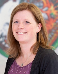
Judith Heidelin
Project manager, LaVision BioTec GmbH
Judith Heidelin finished successfully her diploma thesis in physics in 2005 at Leibniz University of Hannover and her PhD thesis in (Dr. rer. nat.) 2009 at Laser Zentrum Hannover e.V., Germany. After her Postdoc position 2009 – 2011 at École Polytechnique de Montréal, Canada she worked as project manager for laser scanning microscopy customer systems at LaVision BioTec GmbH. In 2016 Judith Heidelin became project manager for grants and collaborative projects and part of the intellectual properties / patents team at LaVision BioTec GmbH. Since 2011 she has managed and accounted different research projects for LaVision BioTec funded by BMBF (TOMOSphere, ELICIT, MoDiaNo), BMWi (LSTM, RETI-IM), and EU (DEEPINSIGHT, FEMSCOPY, DeLIVER).
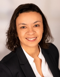
Fernanda Ramos-Gomes
Translational Molecular Imaging, Max-Planck-Institute for Multidisciplinary Sciences
Fernanda Ramos-Gomes is a post-doctoral research fellow in the group of Frauke Alves at the Max-Planck-Institute for Multidisciplinary Sciences in Göttingen. She received her degree in biology from the Federal University of Rio de Janeiro, Brazil. Following her Master in 2006 at the Institute of Biophysics Carlos Chagas Filho in Rio de Janeiro - Brazil, she moved to Germany, where she received her PhD in 2010 at the Göttingen Graduate Center for Neurosciences, Biophysics and Molecular Biosciences (GGNB). In 2011 she joined Frauke Alves’ group of Translational Molecular Imaging. With an excellent background in the handling of biological samples and microscopy techniques, her expertise helps to fill the gap between the preparation of complex tissues and the microscopy imaging scenario. Using different microscopy techniques - high-resolution, multiphoton and light-sheet microscopy - her domain includes deep intra-tumour and intravital imaging, as well as imaging of nonlinear signals produced by tumor structures, drug release in the tumour microenvironment and tracking of nanoparticle-labeled cells. With her expertise she has contributed to several third-party funded applications and has supervised a number of PhD Students. She has a great interest in the oncology field, especially in the processes of how tumor cells interact with components of the extracellular matrix and migrate to form distant metastases.
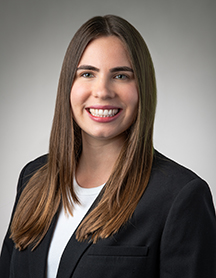
Alexandra Latshaw
PostDoc, University of Geneva
Alexandra earned her Ph.D. in Physics at the University of California, Los Angeles in 2019 characterizing plasma formation in the femtosecond laser breakdown of dense hydrogen gas. She worked at Raytheon Intelligence & Space in El Segundo, California after her Ph.D. as a research scientist where she developed photonic integrated circuit optical phased arrays (PIC OPAs) for high data rate free-space laser communication. She joins the University of Geneva and the FAIR CHARM collaboration as a postdoctoral researcher to implement a new short-wave infrared (SWIR) laser for millimeter-depth, high spatial resolution 3D imaging of pathological tissues.
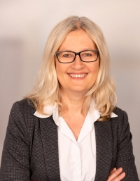
Andrea Markus
Translational Molecular Imaging, Max-Planck-Institute for Multidisciplinary Sciences
Andrea Markus is a senior researcher at the Max-Planck-Institute for Multidisciplinary Sciences, Göttingen. She received her degree in biology from the Johannes Gutenberg University, Mainz. Following her PhD in 1998 at the Helmholtz Center in Munich, she moved to Australia, where she worked in molecular biology of cancer at the University of Sydney. In 2010 she returned to Germany and took up a postdoctoral position in the team of Frauke Alves. She has contributed to the acquisition of a number of EU funded projects and to their implementation with her expertise in diverse imaging technologies, including optical in vivo imaging, high-resolution CT and 2-photon microscopy, for the evaluation and optimization of novel theranostic approaches. She has a particular interest in the question how nanoparticles help to track tumor and immune cells and how they can be exploited for the visualization of cell-cell and cell-stroma interactions, especially in the field of oncology.
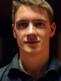
Akos Diosdi
PhD student, Szeged Biological Research Centre (BRC)
Akos Diosdi studied Cell and Molecular Biology at the University of Szeged (Hungary). After obtaining his Bachelor of Science, he joined the Biological Image Analysis and Machine Learning Group (BIOMAG), Synthetic and Systems Biology Unit, Biological Research Centre (BRC, Szeged, Hungary) where he pursued a Master degree in Bioinformatics. In 2019, he received the Discipuli pro Universitate Award for his outstanding performance. Since then, he started his PhD studies under the supervision of Prof. Peter Horvath, and he received the Straub Young Researcher Award for his scientific achievements. His main research fields are: “Light-sheet Microscopy”, “In Vitro 3D Models”, “Imaging Technology”, “Clearing Protocols”, “Computational Biology”. In just 2 years, he collected several contributions in outstanding international conferences and 7 articles published in top scientific journals.
Milvia Alata
Postdoc, KU Leuven
Milvia is a postdoctoral researcher at the Laboratory of Enteric NeuroScience in KU Leuven. She received her degree in exact sciences (physics and mathematics) from the University of Tarapaca in Chile. During her master studies at the Centro de Investigaciones en Optica in Mexico she joined the biophotonics Lab where she worked on the development of a mounting medium for high resolution optical microscopy. She also obtained her PhD at the same institution, working in an interdisciplinary project studying the CNS of a TUBB4A mutant rat using different imaging approaches like MRI, SHG and fluorescence microscopy.
Other contributors, Master & Bachelor Students
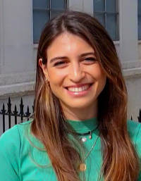
Dora Gibellieri
Master Student, University of Geneva
Dora Gibellieri is completing her Master Thesis at the University of Geneva as part of her Master's programme in Physics. Her research career started during the last year of her Bachelor's degree during which she did an internship at the Institut de Radiophysique IRA/DRM at CHUV (Centre Hospitalier Universitaire Vaudois). The project served as the Bachelor's thesis, granting her a Bachelor's degree in Physics by La Sapienza University in Rome. The following year Dora was granted an internship at CERN, in Geneva, where she participated in a cooperative project in the field of Linac4 transfer-line beam dynamics. The application of several analyzing software and a strong coding base have served her as a solid foundation for the introduction to multi-photon microscopy, which will be the central topic of her Master thesis, as well as her contribution to the FAIR CHARM project.
Marta Margherita Pozzi
Bachelor Student, University of Geneva
Marta is a Bachelor Student from Milan, who came to Geneva within the framework of the Swiss-European mobility programme (SEMP). She completed in November 2022 a Bachelor thesis entitled: Simulation of light transport through biological tissues in the SWIR spectral region

Paulí Figueras
PhD student, University of Geneva
Paulí Figueras Llussà has a Master in Chemistry. He is presently a PhD student in the Department of Applied Physics at the University of Geneva. He is partially involved in FAIR CHARM in aspects related to multiphoton microscopy and the preparation of nanoparticle-labelled samples.
Michel Moret
Research Engineer, University of Geneva
Michel Moret is a research engineer in the Department of Applied Physics at the University of Geneva. His technical and IT support is vessential for the implementation of the SWIM and SLIDE system in Geneva.

