The skeleton of the malaria parasite reveals its secrets
Research teams from UNIGE have discovered that the cytoskeleton of the malaria parasite comprises a vestigial form of an organelle called conoid, initially thought to be absent from this species and which could play a role in host invasion.
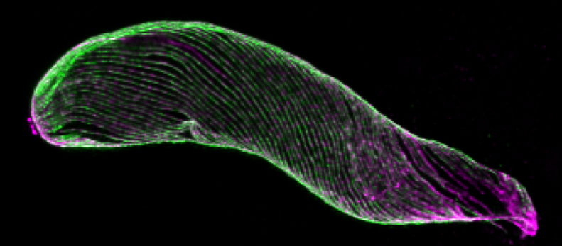
Plasmodium at the ookinete stage viewed by expansion microscopy. The image shows the cytoskeleton of the pathogen following the labelling of tubulin. The conoid is the ring visible at the upper tip of the cell. © UNIGE/HAMEL
Plasmodium is the parasite causing malaria, one of the deadliest parasitic diseases. The parasite requires two hosts —the Anopheles mosquito and the human— to complete its life cycle and goes through different forms at each stage of its life cycle. Transitioning from one form to the next involves a massive reorganisation of the cytoskeleton. Two teams from the University of Geneva (UNIGE) have shed new light on the cytoskeleton organisation in Plasmodium. Their research, published in PLOS Biology, details the organisation of the parasite’s skeleton at an unprecedented scale, adapting a recently developed technique called expansion microscopy. Cells are “inflated” before imaging, providing access to more structural details, at a nanometric scale. The study identifies traces of an organelle called “conoid”, which was thought to be lacking in this species despite its crucial role in host invasion of closely related parasites.
The cytoskeleton, or cell skeleton, consists of a network of several types of filaments, including actin and tubulin. It confers rigidity to the cell, allows the attachment or movement of organelles and molecules inside the cell, as well as cell deformations. As the parasite transitions between developmental stages, its cytoskeleton undergoes repeated, drastic, reorganisations. In particular, Plasmodium needs a very specific cytoskeleton in order to move and penetrate the membrane barriers of its host cells, two processes that are central to the pathogenesis of malaria-causing parasites. “Due to the very small size of Plasmodium—up to 50 times smaller than a human cell—it is a technical challenge to view its cytoskeleton!” begins Eloïse Bertiaux, a researcher at UNIGE and the first author of the study. “That is why we adapted our expansion microscopy protocol, which consists of inflating the biological sample while keeping its original shape, so it can be observed at a resolution that has never been attained before”, continues Virginie Hamel, a researcher at the Department of Cell Biology of the Faculty of Sciences of UNIGE and co-leading the study.
A Vestigial Form of an Organelle
The researchers observed the parasite at the ookinete stage, the form responsible for the invasion of the mosquito midgut, an essential step for the dissemination of malaria. A structure made of tubulin was visible at the tip of the parasite. This structure is similar to a conoid, an organelle involved in host cell invasion, in related Apicomplexa parasites. “The structure observed in Plasmodium seems, however, divergent and reduced compared with the well-described conoid of Toxoplasma, the parasite causing toxoplasmosis. We still need to determine whether this remnant conoid is also important for host cell invasion of Plasmodium”, explains Mathieu Brochet, a professor at the Department of Microbiology and Molecular Medicine of the Faculty of Medicine of UNIGE.
Cytoskeleton Under the Microscope
The discovery of this vestigial conoid highlights the power of expansion microscopy, which can be used to view cytoskeletal structures at the nanoscale without the need for specialised microscopes. Used in combination with electron microscopy and super-resolution microscopy approaches, this method adds molecular details to the available structural information, paving the way for more in-depth studies of the cytoskeleton and its molecular organisation. This will allow us to gain a better understanding of how Plasmodium invades its host cells, a process that is essential for the pathogenesis of this parasite.