Cellular tornadoes sculpt our organs
A team from the UNIGE has demonstrated that cells self-organise to generate forces that model the shapes of our tissues.
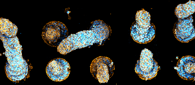
Examples of protrusions formed by confinement of muscle cells on adhesive discs. The disc, slightly larger than the diameter of the protrusion, is visible at the base. On some protrusions, the vortex organization is visible. The twisted shape of other protrusions is also indicative of rotational forces. (c) UNIGE
How are the different shapes of our organs and tissues generated? To answer this question, a team from the University of Geneva (UNIGE), Switzerland, forced muscle cells to spontaneously reproduce simple shapes in vitro. By confining them on adhesion discs, the biochemists and physicists observed that the cells rapidly self-organise by aligning themselves in the same direction. A circular motion is created around a vortex – called a topological defect – which, by orienting the cells, allows them to join forces, deforming the cell monolayer into a protrusion, a structure commonly observed in embryo development. This cylindrical protrusion is maintained by the collective rotational forces of the cells, creating a tornado-like effect. The formation of these cellular tornadoes would therefore constitute a simple mechanism of spontaneous morphogenesis, dictated by the unique properties of multicellular assemblies. These results can be read in the journal Nature Materials.
Our bodies are made up of organs and tissues, each with its peculiar shape. But how do cells manage to form the folds of the intestine or the alveoli of the lungs? Is it possible to reconstitute these shapes in vitro? To answer these questions, biochemists have joined forces with theoretical physicists to test the ability of cellular tissues to spontaneously self-model.
“In theoretical physics, we know that if there are active constraints between cells, then they will order themselves and spontaneously adopt collective behaviours known as ‘emergent’, because they do not exist at the scale of a single cell”, explains Karsten Kruse, professor in the departments of biochemistry and theoretical physics at the UNIGE Faculty of Science. The theory predicts that one of these emerging behaviours is the adoption of particular shapes by a multicellular tissue. It is this hypothesis that we wanted to test in vitro.
To do this, the Geneva team selected human muscle cells that are capable of contracting and whose rod shape allows them to align themselves: “When the cells are placed on a flat surface, they align themselves and form structures similar to a field of wheat where the wind has passed through: there is an overall order with sudden changes in direction at punctual places”, says Aurélien Roux, a professor in the Department of Biochemistry at the UNIGE Faculty of Science. These changes in direction are called ‘topological defects’: they represent the places where the physical forces exerted on the cells are either very weak or, on the contrary, immense.
Topological defects create cellular tornadoes
So what impact do these topological defects have on the shape of the tissue? To understand their role, the interdisciplinary team grew cells on adhesion discs. “This involves confining our muscle cells to a surface surrounded by repulsive molecules that force them to form a circle”, explains Aurélien Roux. The cells quickly start to rotate together to form an ordered spiral. “We can see a spontaneous movement of the cells, like when a crowd is forced to walk around a room and ends up going in the same direction for ease”, he continues.
Thus ordered, only one topological defect remains at the centre of the circle. “We see that the spiral, which concentrates the cellular forces in its centre, accumulates the newly formed cells there by cell division. Thus, the spiral will gradually become a vortex, creating a protrusion in the middle of the disc”, explains Karsten Kruse. And this protrusion can reach up to half a millimetre, which is enormous for a base that is not a hundredth of a millimetre in size. The Geneva team is therefore observing a real little 3D cellular tornado that is spinning around.
Spontaneous cell morphogenesis subject to the laws of physics
The researchers found that the muscle cells spontaneously formed tornado-like structures, which resemble the structures observed in the development of the embryo, such as the fingers or the folds of the intestinal layer. “This spontaneous self-organisation without biochemical regulation could be the initial stage in the formation of protrusions in the embryo”, says Aurélien Roux. The scientists also highlighted that it is indeed the topological defects that control the organisation of cells and determine the shape they will adopt. “Finally, our study shows that cells do not escape the laws of physics but, subjected to the same constraints as all materials, they exploit them to concentrate their forces and create shapes only seen in living organisms”, adds Karsten Kruse.
The researchers will now study simple examples of embryos in order to compare them with theoretical models and in vitro experiments and understand the different possible mechanisms regulating the forces in the embryo.
10 Feb 2022