
Electron Microscopy Facility (PFMU)
Electron Microscopy Facility (PFMU) provides an unprecedented access to study and analyze biological samples at the ultrastructural level with resolution in nanometer scale.
Electron microscopy enables to visualize normal and abnormal cellular and tissue morphology, to analyze the intracellular localization of proteins, lipids and other macromolecules and/or reconstructing the 3D ultrastructure of cells and tissues.
Electron Microscopy Facility is an open door multi-user facility performing most aspects of biological research requiring the use of transmission and/or scanning electron microscopy predominantly applied to cellular electron microscopy (TEM, SEM) using conventional chemical fixation for resin embedded samples for:
- ultramicrotomy and ultra-thin sectioning for TEM analysis,
- cyo-ultramicrotomy and immune-gold labelling by Tokuyasu method,
- volume imaging using focused ion beam and SEM imaging (FIBSEM) for 3D reconstruction of cell or tissues,
- SEM imaging,
- negative staining of macromolecular assemblies,
- bacteriophages,
- viruses and many more.
Thematics
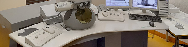
Equipment
- Transmission Electron Microscopes: Tecnai 12G2 and Morgagni (FEI)
- Focused Ion Beam Scanning Electron Microscope (FIBSEM): Helios 660 Nanolab DualBeam FIBSEM (ThermoFisher Scientific)
- Ultramicrotomes: 3x room temperature (Leica Ultracut UCT and Reichert-Jung Ultracut E)
- Cryo-ultramicrotomes: 2x (Leica EMFC7 and UCT-EMFCS)
- Glass knife maker (Leica)
- BAF 060 Freeze-Fracture/Etching System (Bal-Tec)
- Sputter coater for gold and carbon coating: Q150T ES (Quorum)
- Glow-discharge apparatus for negative staining
- Critical-point dryer for SEM dehydration: K850 (Quorum)
- Fully equipped labs for EM specimen preparation
Services
Advisory and consultation services:
- Help to design the experiment, optimal method to prepare the specimen, right choice of the instrument to collect images.
- Specimen preparation for TEM and SEM Services include conventional fixation, dehydration, epon-embedding and sectioning of cells and tissues, cryo-sectioning, immunolabeling, freeze-fracture, critical point drying of specimen, metal coating, negative staining, volumeEM by FIBSEM, etc.
- Access to the transmission electron microscopes and ancillary equipment Independent access to electron microscopes and ancillary equipment is possible after adequate training.
- Training sessions to operate the instruments properly and efficiently Since most new users are not familiar with the instruments, the PFMU staff will train users on a one-to-one basis. When the instructor considers a person capable of working by her/himself, she/he will be allowed to use the facility equipment.
Address
Centre Médical Universitaire (CMU)
1 rue Michel-Servet 1
1211 Genève 4