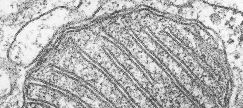
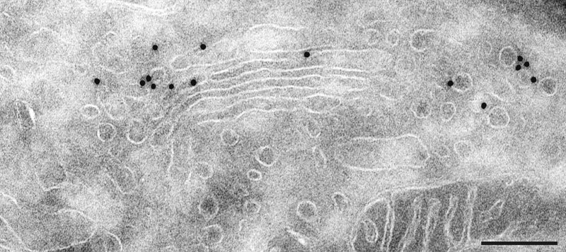
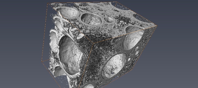
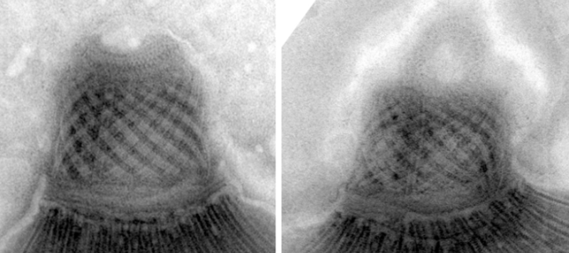
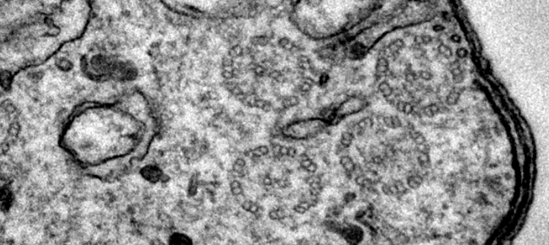
Electron microscopy (EM) provides unprecedented access to study and analyze biological samples at the ultrastructural level, with resolution in nanometers scale.
EM is notably suitable for:
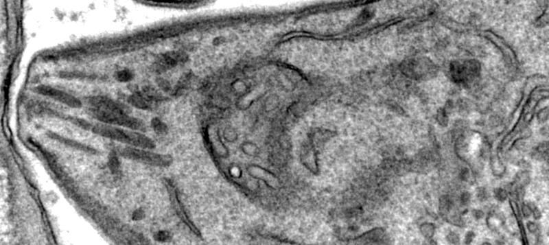
- visualizing normal and abnormal cellular and tissue morphology
- analyzing the intracellular localization of proteins, lipids and other macromolecules
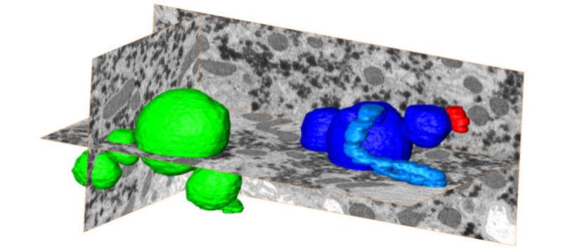
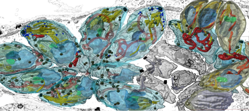
- reconstructing the 3D ultrastructure of cells and tissues
The Electron Microscopy Facility (Pôle Facultaire de Microscopie Ultrastructurale (PFMU)) at the Medical Faculty of the University of Geneva:
- is an open door multi-user facility performing most aspects of biological research that require the use of scanning and/or transmission electron microscopy (SEM/TEM).
- anyone in the University research community who needs to use the PFMU facilities is welcome to have an access to facility infrastructure
- PFMU is also open to users from other universities and from industry.
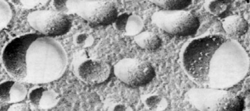
- PFMU specializes for cellular electron microscopy (TEM and SEM) using conventional chemical fixation for:
- ultramicrotomy and ultra-thin sectioning for TEM analysis
- cryo-ultramicrotomy and immuno-gold labelling
- volume imaging using focused ion beam and SEM imaging (FIBSEM) for 3D reconstruction of the cells or tissues
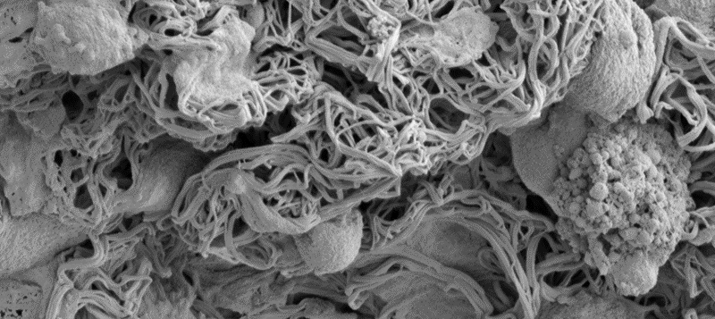
- SEM imaging
- negative staining
- ...
To cover part of the costs and to be able to maintain the equipment, fees to use the equipment and fees for specimen preparation are charged to the users.
Initial contact
A prospective user should contact a member of the PFMU staff (see Contacts) and determine if the instruments in the facility are suitable for the proposed research.