Molecular Imaging: Micro PET/SPECT/CT (CIBM)
Positron Emission Tomography (PET) and Single Photon Emission Computed Tomography (SPECT) are in vivo molecular imaging technologies that use the emission of high-energy gamma rays to visualize and measure biological processes within living animals. Drug candidates, cells or biomarkers can be labeled with a positron-emitting radioisotope (e.g., 18F, 11C) or gamma-emitting radioisotope (e.g., 99mTc, 111In, 131I) and then injected into a study subject.
PET is extremely sensitive, enabling quality images even with very small amounts of injected activity. The penetration capabilities of the gamma rays allow imaging at virtually unlimited depth.
Micro-SPECT shares the penetration capabilities of PET, enabling imaging at virtually unlimited depth. The gamma-emitting radioisotopes used in SPECT are cheaper and have longer half-lives than PET isotopes, rendering SPECT a more practical and economical imaging modality. However, because most of the gamma events do not pass through the pinhole(s), micro-SPECT has more limited sensitivity than PET, leading to longer scan times or larger dose requirements at a constant level of resolution.
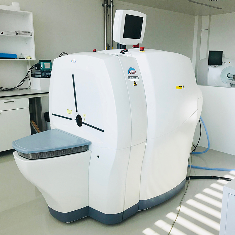
PET/SPECT/CT scanner