Chemotherapy-driven intestinal dysbiosis and indole-3-propionic acid rewire myelopoiesis to promote a metastasis-refractory state
Colorectal cancer (CRC) is the second leading cause of cancer-related death worldwide. While early-stage disease can often be cured by surgery, most patients have microsatellite stable (MSS) tumors that do not respond to immune checkpoint inhibitors. For high-risk stage II and stage III CRC, adjuvant chemotherapy with fluoropyrimidines and oxaliplatin reduces recurrence and improves survival. However, 30–50% of patients still develop distant metastases, most commonly in the liver.
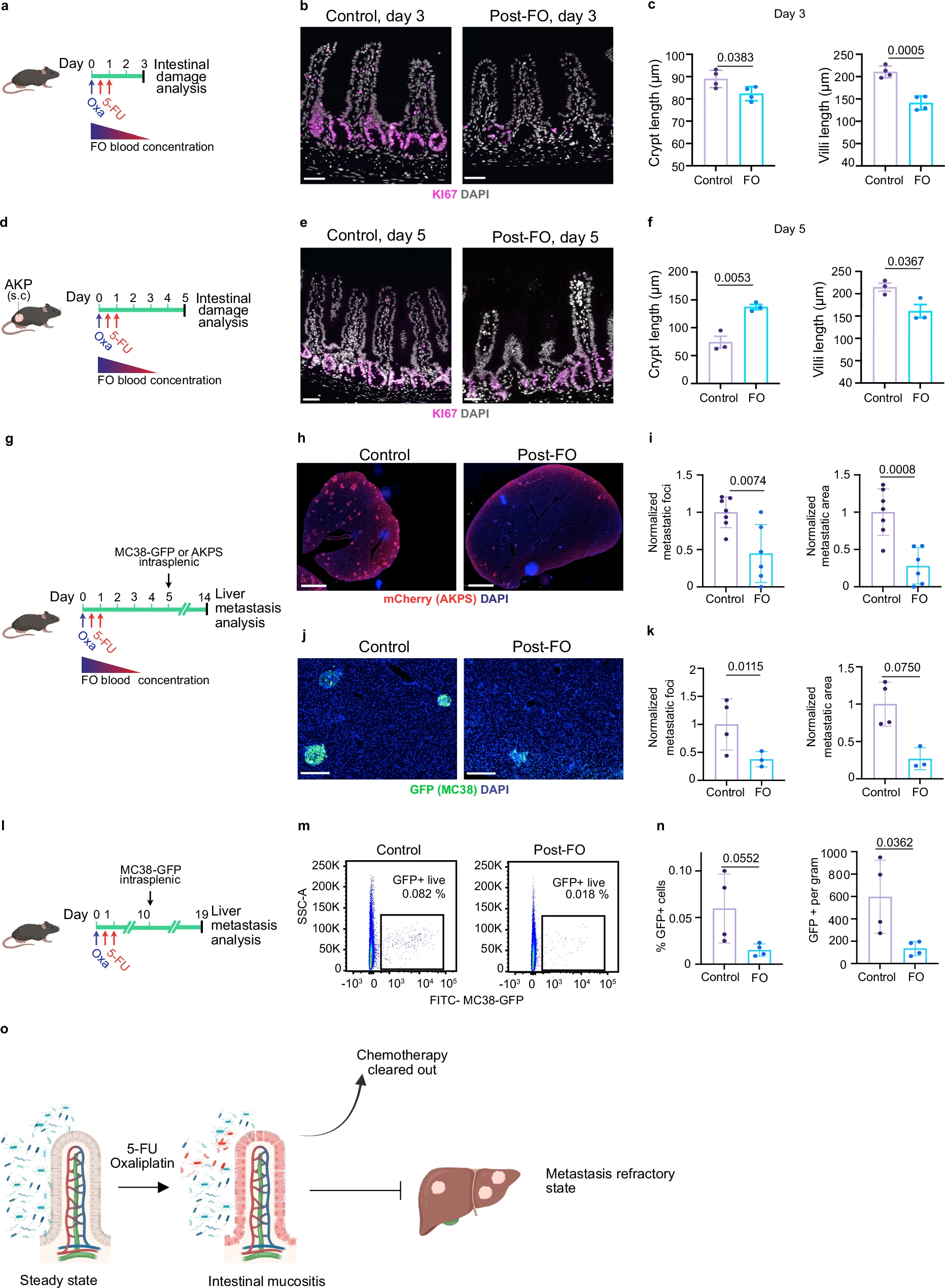
a Scheme of the experiment. Wild-type mice received 5-FU+oxaliplatin (FO) or PBS (control); intestines were harvested 3 days later. b FO reduced proliferation of intestinal crypt cells. KI67 (magenta) and DAPI (grey). Scale bars: 100 μm. c Quantification of crypt (left) and villi (right) length 3 days post-FO. n = 4 per condition. d Scheme of the experiment. AKP organoids were injected subcutaneously and FO treatment was initiated when tumor reached 130 mm3. Tissues were harvested 5 days after FO. e Hyperproliferative crypt cells five days after FO. KI67 (magenta) and DAPI (grey). Scale bars: 100 μm. f Quantification of crypt (left) and villi (right) length five days post-FO. n = 3 per condition. g Scheme of the experiment. AKPS-mCherry or MC38-GFP cells were injected intrasplenically 5 days after FO preconditioning, when active form of FO has been cleared-out. h FO preconditioning reduces liver metastasis formation in AKPS model. Staining for tumor cells (mCherry, red) and DNA (blue). Scale bar: 2 mm. i Quantification of AKPS metastasis area and number per liver area normalized to control mean. Data are from two independent experiments. Control, n = 7. FO, n = 6. j FO preconditioning reduces liver metastasis formation in MC38-GFP model. Staining for GFP (green) and DNA (blue). Scale bar: 200 μm. k Quantification of MC38-GFP metastasis area and number per liver area normalized to control mean. Control, n = 4. FO, n = 3. l Scheme of the experiment. MC38-GFP cells were injected intrasplenically 10 days after FO preconditioning. m FO chemomemory is lasting. Representative FACS plots of GFP+ cancer cells in livers of control and FO pre-conditioned mice. n Quantification of percentage and count of GFP positive cancer cells in the indicated conditions. n = 4 per condition. o Chemotherapy exposure exerts a lasting antimetastatic effect on CRC liver metastasis. Data are shown as mean ± SD and analyzed using two-tailed unpaired Student’s t- test (c, f, i, k, n).
Growing evidence shows that the gut microbiota influences the effectiveness of chemotherapy. In preclinical models, microbial sensing is required for optimal responses to oxaliplatin, and chemotherapy-induced microbial translocation can stimulate anti-tumor CD4⁺ T cell responses. Clinically, patients often receive antibiotics around the time of chemotherapy, and a large retrospective study found that antibiotic use was associated with worse disease-free survival in CRC. Despite this, the mechanisms linking chemotherapy, microbiota changes, and metastatic relapse remain poorly understood.
Here is shown that standard chemotherapy with 5-FU and oxaliplatin induces a long-lasting, metastasis-resistant state by durably reshaping the gut microbiota. This remodeling increases production of the microbiota-derived tryptophan metabolite indole-3-propionic acid (IPA). IPA acts primarily on myeloid progenitors in the bone marrow, promoting macrophage differentiation while reducing immunosuppressive Ly6C^high^CCR2⁺ monocytes. This shift enhances anti-tumor CD4⁺ T cell responses and limits CRC liver metastasis.
These findings are supported by patient data showing that circulating IPA levels increase after chemotherapy in a subset of CRC patients and inversely correlate with monocyte abundance. Together, our results reveal a previously unrecognized systemic effect of chemotherapy mediated by the gut microbiota and identify IPA as a potential therapeutic strategy to counteract immunosuppression and metastasis.