The role of Panx1 in cardiac ischaemia & reperfusion
© The Author(s) 2023. Published by Oxford University Press on behalf of the European Society of Cardiology
SUMMARY
No effective therapy is available in clinics to protect the heart from ischaemia/reperfusion (I/R) injury. Endothelial cells are activated after I/R, which may drive the inflammatory response by releasing ATP through pannexin1 (Panx1) channels. Here, the authors, led by GCIR Professor Brenda Kwak, investigated the role of Panx1 in cardiac I/R.
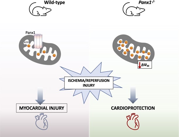
Panx1 was found in cardiac endothelial cells, neutrophils, and cardiomyocytes. After in vivo I/R, serum Troponin-I, and infarct size were less pronounced in Panx1-/- mice, but leukocyte infiltration in the infarct area was similar between Panx1-/- and wild-type mice. Serum Troponin-I and infarct size were not different between mice with neutrophil-specific deletion of Panx1 and Panx1fl/fl mice, suggesting that cardioprotection by Panx1 deletion rather involved cardiomyocytes than the inflammatory response. Physiological cardiac function in wild-type and Panx1-/- hearts was similar. The time to onset of contracture and time to maximal contracture were delayed in Panx1-/- hearts, suggesting reduced sensitivity of these hearts to ischaemic injury. Moreover, Panx1-/- hearts showed better recovery of left ventricle developed pressure, cardiac contractility, and relaxation after I/R. Ischaemic preconditioning failed to confer further protection in Panx1-/- hearts. Panx1 was found in subsarcolemmal mitochondria (SSM). SSM in WT or Panx1-/- hearts showed no differences in morphology. The function of the mitochondrial permeability transition pore and production of reactive oxygen species in SSM was not affected, but mitochondrial respiration was reduced in Panx1-/- SSM. Finally, Panx1-/- cardiomyocytes had a decreased mitochondrial membrane potential and an increased mitochondrial ATP content.
Panx1-/- mice display decreased sensitivity to cardiac I/R injury, resulting in smaller infarcts and improved recovery of left ventricular function. This cardioprotective effect of Panx1 deletion seems to involve cardiac mitochondria rather than a reduced inflammatory response. Thus, Panx1 may represent a new target for controlling cardiac reperfusion damage.
Full article: https://doi.org/10.1093/cvr/cvad120
This work was supported by the Swiss National Science Foundation, the Swiss Life Foundation and the Carlos et Elsie De Reuter Foundation.
Why is it important?
Ischemic heart disease is the leading cause of death worldwide. An occluded coronary artery causes an inadequate blood supply to the heart and leads to a heart attack. Reperfusion injury is the tissue damage caused when the blood supply returns to the tissue (re + perfusion) after a period of ischemia. The massive cell death in the heart that occurs in the early stages of reperfusion can lead in some cases to heart failure. New cardioprotective strategies need to be defined in order to treat ischemia-reperfusion (I/R) injury.
The authors of this article are worldwide experts on pannexin (Panx) channels, which are plasma membrane channels that mediate cell signalling. Panx1 channels have been suggested to play a role in the regulation of inflammation and repair after ischemic injury.
The authors used a mouse model, ex vivo cardiac function assays, measured mitochondrial respiration in cells and isolated mitochondria and performed confocal and electron microscopy to elucidate whether Panx1 channels contribute to cardiac I/R injury. They found that Panx1 deletion confers cardioprotection against I/R by increasing mitochondrial ATP content resulting in a decreased sensitivity to cardiac ischemia and a better recovery of cardiac function after the insult. Given that mitochondria have been recognized to be primarily responsible for cell death and survival in cardiomyocytes, Panx1 could represent a novel target for cardioprotection.
24 Aug 2023