Equipment specification
Our bioimaging facility hosts state-of-the-art equipment with:
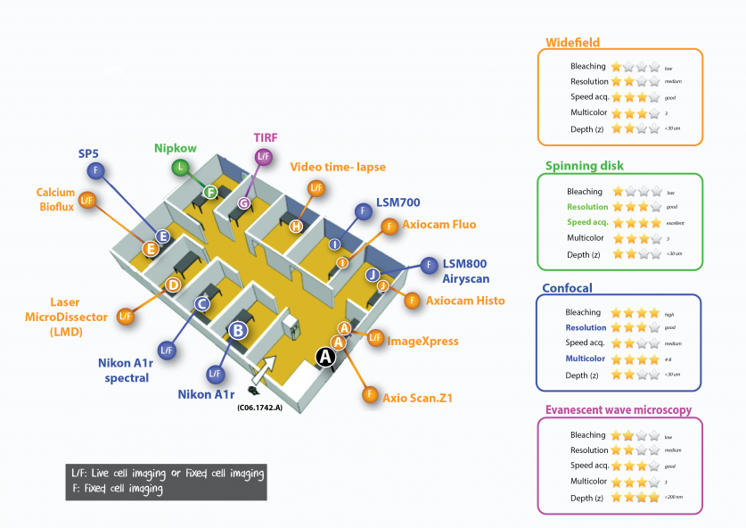
- 15 imaging systems (1 spinning disk confocal microscope (Nipkow), 5 confocal laser scanning microscopes (CLSM), 2 slide scanners and 7 widefield fluorescence microscopes including 1 TIRF evanescent wave microscope, 1 laser microdissector, 2 slide scanners and 1 plate scanner/ImageXpress system) that provide a wide range of applications such as cell membrane visualization, laser microdissection/isolation, colocalization analysis, F-techniques (FRAP, FRET), multi-dimensional time-lapse recordings, intravital imaging and high throughput imaging.
The Olympus VS120 in ROOM C06.1533.A (ex-room 6008), otherwise all other of microscopes are located in ROOM C06.1742.A (ex-room 6021):
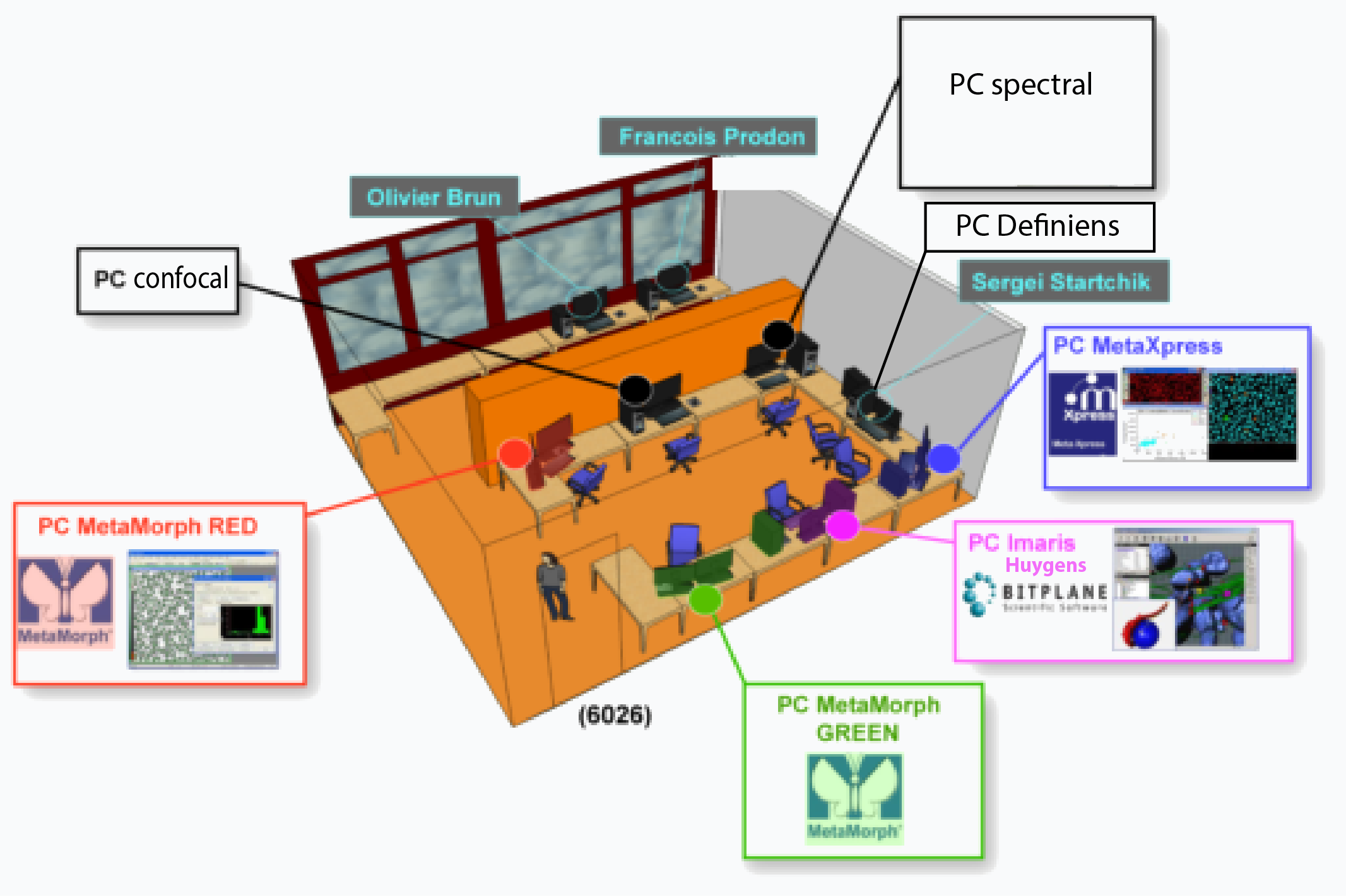
- 7 PC workstations for quantitative analysis and image treatments (MetaMorph, Imaris, Huygens, NIS Nikon, ZEN Zeiss, ImageJ/ Fiji, etc...). These PC are located just in front the main microscope ROOM C06.1742.A (ex-room 6021) in ROOM C06.1542A (ex-room 6026):